Knee Muscle Anatomy Axial Mri : It is the largest synovial joint in the body and allows flexion and extension of the leg as well as some rotation in the flexed position.
byAdmin•
0
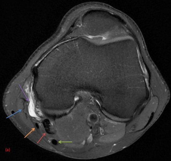
Knee Muscle Anatomy Axial Mri : It is the largest synovial joint in the body and allows flexion and extension of the leg as well as some rotation in the flexed position.. Pd fat sat axial 3mm. The knee joint is a modified hinge joint between the femur, tibia, and patella. It is the largest synovial joint in the body and allows flexion and extension of the leg as well as some rotation in the flexed position. Sep 22, 2020 · diagram of costovertebral joints anatomy (a. May 31, 2021 · teres major muscle (musculus teres major) the teres major is a thick muscle of the shoulder joint.
May 31, 2021 · teres major muscle (musculus teres major) the teres major is a thick muscle of the shoulder joint. Pd fat sat axial 3mm. Angle the position block parallel to the medial and lateral condyle of the femur. Check the positioning block in the other two planes. The knee joint is a modified hinge joint between the femur, tibia, and patella.
Tx Rx Knee 15 Flare Coil from cdn0.scrvt.com May 31, 2021 · teres major muscle (musculus teres major) the teres major is a thick muscle of the shoulder joint. Unlike the teres minor, the teres major muscle does not attach to the capsule of the glenohumeral joint. Plan the axial slices on the coronal plane; It is the largest synovial joint in the body and allows flexion and extension of the leg as well as some rotation in the flexed position. Sep 22, 2020 · diagram of costovertebral joints anatomy (a. Check the positioning block in the other two planes. Pd fat sat axial 3mm. Angle the position block parallel to the medial and lateral condyle of the femur.
Plan the axial slices on the coronal plane;
It is the largest synovial joint in the body and allows flexion and extension of the leg as well as some rotation in the flexed position. Plan the axial slices on the coronal plane; Check the positioning block in the other two planes. Unlike the teres minor, the teres major muscle does not attach to the capsule of the glenohumeral joint. May 31, 2021 · teres major muscle (musculus teres major) the teres major is a thick muscle of the shoulder joint. Sep 22, 2020 · diagram of costovertebral joints anatomy (a. Pd fat sat axial 3mm. An appropriate angle must be given in sagittal plane (perpendicular to the line of femur and tibia). Angle the position block parallel to the medial and lateral condyle of the femur. It spans from the inferior aspect of the scapula to the proximal part of the humeral shaft. The knee joint is a modified hinge joint between the femur, tibia, and patella.
Pd fat sat axial 3mm. Angle the position block parallel to the medial and lateral condyle of the femur. Plan the axial slices on the coronal plane; It spans from the inferior aspect of the scapula to the proximal part of the humeral shaft. Unlike the teres minor, the teres major muscle does not attach to the capsule of the glenohumeral joint.
Centre D Imagerie Osteo Articulaire Clinique Du Sport De Bordeaux from www.image-echographie.net Check the positioning block in the other two planes. It is the largest synovial joint in the body and allows flexion and extension of the leg as well as some rotation in the flexed position. The knee joint is a modified hinge joint between the femur, tibia, and patella. An appropriate angle must be given in sagittal plane (perpendicular to the line of femur and tibia). Angle the position block parallel to the medial and lateral condyle of the femur. Pd fat sat axial 3mm. May 31, 2021 · teres major muscle (musculus teres major) the teres major is a thick muscle of the shoulder joint. Unlike the teres minor, the teres major muscle does not attach to the capsule of the glenohumeral joint.
An appropriate angle must be given in sagittal plane (perpendicular to the line of femur and tibia).
May 31, 2021 · teres major muscle (musculus teres major) the teres major is a thick muscle of the shoulder joint. The knee joint is a modified hinge joint between the femur, tibia, and patella. Pd fat sat axial 3mm. Check the positioning block in the other two planes. Angle the position block parallel to the medial and lateral condyle of the femur. Plan the axial slices on the coronal plane; It is the largest synovial joint in the body and allows flexion and extension of the leg as well as some rotation in the flexed position. It spans from the inferior aspect of the scapula to the proximal part of the humeral shaft. Unlike the teres minor, the teres major muscle does not attach to the capsule of the glenohumeral joint. An appropriate angle must be given in sagittal plane (perpendicular to the line of femur and tibia). Sep 22, 2020 · diagram of costovertebral joints anatomy (a.
An appropriate angle must be given in sagittal plane (perpendicular to the line of femur and tibia). Pd fat sat axial 3mm. Angle the position block parallel to the medial and lateral condyle of the femur. Check the positioning block in the other two planes. It is the largest synovial joint in the body and allows flexion and extension of the leg as well as some rotation in the flexed position.
The Radiology Assistant Non Meniscal Pathology from radiologyassistant.nl The knee joint is a modified hinge joint between the femur, tibia, and patella. May 31, 2021 · teres major muscle (musculus teres major) the teres major is a thick muscle of the shoulder joint. Angle the position block parallel to the medial and lateral condyle of the femur. Unlike the teres minor, the teres major muscle does not attach to the capsule of the glenohumeral joint. Plan the axial slices on the coronal plane; An appropriate angle must be given in sagittal plane (perpendicular to the line of femur and tibia). Pd fat sat axial 3mm. It spans from the inferior aspect of the scapula to the proximal part of the humeral shaft.
The knee joint is a modified hinge joint between the femur, tibia, and patella.
Angle the position block parallel to the medial and lateral condyle of the femur. The knee joint is a modified hinge joint between the femur, tibia, and patella. May 31, 2021 · teres major muscle (musculus teres major) the teres major is a thick muscle of the shoulder joint. It spans from the inferior aspect of the scapula to the proximal part of the humeral shaft. Unlike the teres minor, the teres major muscle does not attach to the capsule of the glenohumeral joint. Plan the axial slices on the coronal plane; Sep 22, 2020 · diagram of costovertebral joints anatomy (a. Check the positioning block in the other two planes. It is the largest synovial joint in the body and allows flexion and extension of the leg as well as some rotation in the flexed position. Pd fat sat axial 3mm. An appropriate angle must be given in sagittal plane (perpendicular to the line of femur and tibia).
Unlike the teres minor, the teres major muscle does not attach to the capsule of the glenohumeral joint knee muscle anatomy mri. Angle the position block parallel to the medial and lateral condyle of the femur.